
Talk with your doctor about your own report. The notes below are general, and they are not a reading of your scan. When you want help preparing questions, use the fibroid visit checklist or the treatment options page, since neither page reads a report.
Where fibroids can sit
The uterus is a muscle with a lining on the inside and a smooth outer surface. A fibroid can sit in the inner space, in the wall, or on the outside, and it can also hang from a stalk or sit in the cervix.
FIGO types 0 to 8
Doctors use numbers called FIGO types to say where a fibroid sits compared with the lining and the outer surface. The numbers run from 0 to 8.2, 6 Your report may use these numbers, or it may use words only.5
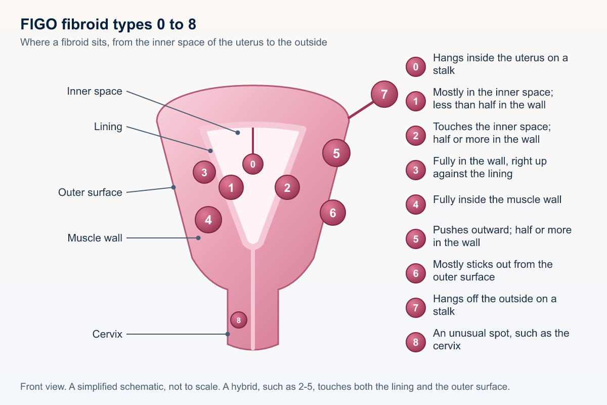
| Type | Where it sits |
|---|---|
| 0 | Hangs inside the uterus on a stalk. It is fully in the inner space. |
| 1 | Mostly bulges into the inner space. Less than half sits in the wall. |
| 2 | Touches the inner space, but half or more sits in the wall. |
| 3 | Fully in the wall, right up against the lining. |
| 4 | Fully inside the muscle wall. |
| 5 | Pushes outward, but half or more is in the wall. |
| 6 | Mostly sticks out from the outer surface. |
| 7 | Hangs off the outside of the uterus on a stalk. |
| 8 | In an unusual spot, such as the cervix, or not attached to the uterus. |
| Hybrid, such as 2-5 | Touches both the lining and the outer surface. It spans the whole wall. The first number is the lining side. The second number is the outer side. |
A 2018 update kept this same set of numbers, including hybrids.6
Why the type number matters
The type helps your doctors think about which treatments may fit. Whether a fibroid sits in the inner space, in the wall, or on the outside can steer that talk. A fibroid on a stalk (type 0 inside, type 7 outside) may get a closer look before embolization, because the stalk width can matter. Your doctor can explain which treatments fit each type.2, 4, 6
Words you may see on the report
The circled numbers match the fictional sample below, and a report may use the medical word, the plain word, or both.
-
Term 1. T1 and T2 pictures
These are two kinds of MRI pictures, each showing tissue in a different shade of gray, and doctors compare them to tell what something is made of. A report may say “T1-weighted” or “T2-weighted.”5
-
Term 2. T2 dark (low signal)
A typical fibroid looks dark on T2 pictures because it is dense muscle, and this is the usual look. A report may say “low signal” or “hypointense.”5
-
Term 3. T2 bright or patchy
Brighter or patchy areas can mean more fluid or a change inside the fibroid, which is covered under degeneration below, and most of the time this is still not cancer. Sometimes the doctor looks more closely, and a report may say “high signal” or “heterogeneous.”5
-
Term 4. T1 bright (high signal)
Brightness on T1 can point to old blood or fat inside a fibroid, and old blood fits a change called red degeneration.5
-
Term 5. Contrast enhancement
A dye is given through an IV, parts with a blood supply light up, and doctors call that enhancement. It shows which fibroids are alive and fed by blood.4, 5
-
Term 6. Non-enhancement
The fibroid, or part of it, does not light up, so it has little or no blood supply, and a report may say “non-enhancing,” “devascularized,” or “infarcted.” Before treatment it may have already died off, and after embolization this is the expected look.4, 5
-
Term 7. Degeneration
A fibroid can change inside over time, often when it outgrows its blood supply, and these changes are common and are usually not cancer. Hyaline means firm, glassy tissue, cystic means fluid pockets, and myxoid means jelly-like material. Red, also called hemorrhagic, means bleeding inside, and it can happen in pregnancy and can hurt. Calcific means calcium makes it hard, often after menopause.5
-
Term 8. Submucosal
The fibroid pushes into the inner space of the uterus, where the lining is.2, 5, 6
-
Term 9. Intramural
-
Term 10. Subserosal
-
Term 11. Pedunculated (stalk)
The fibroid is attached by a stalk, which can be inside the uterus or outside it, and the report may say how wide the stalk is.4, 5
-
Term 12. Cervical
The fibroid sits in the cervix, the lower neck of the uterus.2, 6
-
Term 13. Size and volume
Size is three numbers, length, width, and depth, in centimeters, and the report may also list a volume. The dominant fibroid is the largest one, or the one that matters most for the visit.5
-
Term 14. Adenomyosis and the junctional zone
Adenomyosis is a separate condition in which lining-type tissue grows into the muscle wall. On MRI, an inner band of that wall, called the junctional zone, can look thick. Reports sometimes list a thickness. A junctional zone of 12 mm or more has often been used as a sign of adenomyosis, but thickness alone is not a sure test. Doctors also look at its shape and for tiny cysts in the wall.5, 8
-
Term 15. Ovaries
The report usually describes the ovaries, and simple cysts are common and often normal in people who still have periods. “Adnexa” means the ovaries and the tissue beside the uterus.5
-
Term 16. Cannot exclude leiomyosarcoma
Rarely, a growth that looks like a fibroid is a cancer called leiomyosarcoma. MRI describes fibroids well, but no scan can rule this out completely. When a report says this, the reader is usually being careful, and it does not mean cancer was found. Your care team will talk with you about any next step.4, 5
-
Term 17. Enhancement, avid, or heterogeneous enhancement
These words describe how much and how evenly a part lights up with contrast dye.5
-
Term 18. Diffusion-weighted imaging (DWI) and ADC
These are pictures of how freely water moves in tissue. They help doctors characterize what they see.5
-
Term 19. Myometrium, endometrium, and serosa
The muscle wall, the lining, and the outer surface of the uterus.5
-
Term 20. Fundus, body, lower segment, anteverted, and retroverted
The top, middle, and lower part of the uterus, and whether it tilts forward or back.5
-
Term 21. Mass effect and hydronephrosis
Pressing on nearby organs, and swelling of a kidney when urine flow is blocked.5 If your report uses these words, ask about them promptly.
-
Term 22. Infarcted or devascularized
Without blood flow.5
-
Term 23. Adenomyoma
A lump of adenomyosis, which can look like a fibroid on imaging.5
-
Term 24. Collateral or ovarian arterial supply
A second artery route to the uterus, sometimes from the ovary.5
What enhancement means for embolization
Embolization works by blocking the small arteries that feed a fibroid, so the fibroid shrinks. On the MRI, the contrast dye shows which fibroids have that blood supply. A fibroid that lights up (enhances) has blood flow to block. A fibroid that does not light up (non-enhancing) already has little or no blood supply. Blocking its arteries is unlikely to change it much, so it may not shrink or ease symptoms after embolization. After embolization, non-enhancing is the look doctors hope to see, because it means the treatment cut off the blood flow.5, 7, 9
When a fibroid changes inside
As fibroids grow, some outgrow their blood supply and change inside. This is called degeneration. Reports may use words such as hyaline, cystic, myxoid, red degeneration or hemorrhagic change, or calcification to describe those changes. Your doctor can explain what the words on your report mean.5
Things that can look like fibroids
Adenomyosis is when lining-type tissue grows into the muscle wall. It can cause heavy, painful periods, and it can occur along with fibroids. Signs on MRI include a thick or uneven inner band (the junctional zone) and tiny bright spots in the wall.5 A thickness of about 12 mm or more is often mentioned, but thickness alone is not a sure sign.8 An adenomyoma is a lump of adenomyosis. It can look like a fibroid, but it usually has fuzzy edges instead of a smooth border. Other look-alikes include a polyp in the lining and a growth from the ovary sitting next to the uterus. MRI helps tell these apart, and that can change which treatments make sense.5
A short note on leiomyosarcoma
Rarely, a growth that looks like a fibroid is a cancer called leiomyosarcoma. MRI describes fibroids well, but no scan can rule this out completely. If a report says it cannot be excluded, the reader is usually being careful, and it does not mean cancer was found. Your care team will talk with you about any next step.4, 5, 10
Studies show MRI can help doctors notice features that are more worrying, especially when several features appear together. But even the best scans still miss some cancers and sometimes flag harmless fibroids. That is why no report can promise a fibroid is not cancer. Your care team weighs the scan together with your age, symptoms, and history.10, 12, 14, 15
A fictional sample report
The sample below is made up so you can see how those numbered words look together, and it is not a real person or a real scan. Do not use it to judge your own report.
Exam
MRI pelvis without and with IV contrast (FICTIONAL)
Patient: “Sample Patient”, age 4X · Date: none · MRN: none
Clinical history: Heavy menstrual bleeding and pelvic pressure. Evaluation before possible uterine fibroid embolization.
Technique
Multiplanar T2-weighted ① and axial T1-weighted ① images of the pelvis, including T1 fat-saturated images, diffusion-weighted imaging, and dynamic post-contrast T1 images after IV gadolinium ⑤. Limited MR angiography.
Findings
Uterus: Anteverted, enlarged, measuring 13.2 × 9.8 × 10.4 cm (volume about 704 mL) ⑬. Multiple fibroids, at least 6.
1. Dominant fibroid: intramural ⑨, posterior fundus, 7.1 × 6.4 × 6.8 cm (about 162 mL) ⑬. Mostly T2 hypointense ② with a central T2 hyperintense, non-enhancing area ③⑥ consistent with cystic/hyaline degeneration ⑦. The rest enhances avidly ⑤.
2. Submucosal fibroid ⑧, anterior body, FIGO type 1, 2.4 cm, T2 hypointense ②, enhancing ⑤. Distorts the endometrial cavity.
3. Subserosal pedunculated fibroid ⑩⑪, right lateral, FIGO type 7, 3.0 cm, stalk width 1.1 cm ⑪. Enhancing.
4. Intramural/subserosal fibroid, anterior, FIGO type 2-5 (hybrid), 3.6 cm, enhancing.
5. Small fibroid with intrinsic T1 hyperintensity ④ and no enhancement ⑥, likely prior hemorrhagic (red) degeneration ⑦, 1.8 cm.
6. Cervical fibroid ⑫, posterior lip, 1.2 cm.
Junctional zone: up to 8 mm. No MRI features of adenomyosis ⑭.
Endometrium: 6 mm, displaced by fibroid 2.
Ovaries: Right ovary normal. Left ovary with a 2.1 cm simple-appearing cyst, likely physiologic ⑮.
Vessels: Uterine arteries enlarged on both sides. No definite enlarged ovarian arterial supply to the fibroids is seen.
Other: No free fluid. No enlarged lymph nodes. Mild fullness of the right renal collecting system, likely from mass effect.
Atypical features: none of the dominant lesions shows features that raise particular concern. As with all imaging, MRI cannot completely exclude leiomyosarcoma ⑯.
Impression (fictional)
1. Enlarged fibroid uterus with at least 6 fibroids. The dominant one is a 7.1 cm posterior intramural fibroid with partial degeneration. Most fibroids enhance ⑤.
2. Submucosal FIGO type 1 fibroid distorting the cavity ⑧.
3. Pedunculated subserosal fibroid with a 1.1 cm stalk ⑪.
4. No MRI evidence of adenomyosis ⑭.
5. Small left ovarian cyst, likely physiologic ⑮.
If your report is from an ultrasound
Fibroids are often first found on a pelvic exam or an ultrasound. An ultrasound uses sound waves to make a picture of the uterus and the other organs in the pelvis.16 Before embolization, an MRI or an ultrasound of the uterus is used to check the size, number, and location of the fibroids.17
An ultrasound report uses many of the same words as an MRI report, such as submucosal, intramural, and subserosal, and it may give a FIGO type. Those words are explained above. Ultrasound is often the first test, and an MRI can add detail about complex anatomy, adenomyosis, or planning before a procedure. If you only had an ultrasound, you can ask what an MRI would answer that the ultrasound did not.
More words from ultrasound reports
- Enlarged uterus. The report says the uterus is bigger than expected. You can ask what is making it larger.
- Vascularity. This means blood flow. A report may describe how much blood flow a fibroid has, and “atypical vascularity” is wording to ask about, not a verdict.
- Sonohysterography. Fluid is placed in the uterus through the cervix while ultrasound pictures are taken of the inside of the uterus.16
- Hysteroscopy. A thin scope goes in through the vagina and the cervix, so the doctor can see fibroids inside the uterine cavity.16
- Obstruction. Something is blocking the flow of urine. See the next box.
Questions to ask
- Is kidney function at risk, and has it been checked?
- Do I need urology input, labs, or more imaging?
- Would sonohysterography, hysteroscopy, or an MRI clarify anything on my report?
- Is the blood flow wording expected, or does it need follow-up or a closer look?
Worth knowing: Radiologists and clinicians may read words like “atypical vascularity” or “suspicious features” in different ways. If wording on your report raises a concern, ask your clinician to explain it. You can also ask whether a radiologist who focuses on the female pelvis (gyn-radiology) can give a second read, rather than assuming the wording is routine.
Why MRI matters before UFE
If you and your doctor are talking about uterine fibroid embolization, MRI is often part of the plan. You may also hear the procedure called uterine artery embolization, or UFE or UAE for short.
For known fibroids, when the visit is about planning treatment, two tests are usually appropriate. One is an MRI of the pelvis with and without IV contrast dye. The other is a pelvic ultrasound. CT is usually not the test used for this.11
An updated radiology guideline on treating fibroids (2023) discusses embolization, MRI-guided focused ultrasound, and surgery to remove fibroids.13
Before embolization, ultrasound, MRI, or both help confirm the diagnosis. They also help the team decide whether the procedure is a fit. That guidance also covers fibroids on a stalk, adenomyosis, medicines already tried, and wishes about a future pregnancy.4, 10
MRI helps the team plan embolization in several ways:
- Number, size, and location of every fibroid. Ultrasound can miss small or deep ones.
- Uterine volume: a starting number to compare with later scans.
- Blood supply: which fibroids enhance and are likely to respond.
- Extra arteries from the ovary: an MRI artery scan can hint that an ovarian artery also feeds the fibroids. This can change the procedure plan.3
- Other causes of symptoms: adenomyosis, polyps, ovarian masses, or rarely a worrying feature.
- Changed plans: in studies, findings on the pre-procedure MRI changed or added to the plan for some patients.1
A full report often includes the number, the size, the location, and the picture signal. It also notes degeneration, whether the fibroids enhance, adenomyosis, the junctional zone, and other findings. Some scans add a look at the arteries.4, 5, 11
Features a doctor may care about
These are general reasons, not a verdict on your MRI.4, 5
| Feature | Why a doctor may care |
|---|---|
| Number | Many fibroids can be treated at one time with embolization, and the count helps compare options. |
| Size | Very large fibroids, or a very large uterus, may respond in a different way, and size is also the starting point for later checks. |
| Location | Fibroids mostly in the cavity, types 0 and 1, may pass or may be removed through the cervix, and location also ties to symptoms. |
| Stalk | A thin stalk has raised concern that a fibroid could come loose after embolization, but selected fibroids on a stalk can still be treated, so ask what your measurement means for you.4 |
| Enhancement | Fibroids with a good blood supply are the ones embolization targets, and a fibroid that does not light up may not benefit. In retrospective single-center studies, fibroids that lit up well and those inside the cavity tended to shrink more after embolization. Results vary from person to person.5, 7, 9 |
| Adenomyosis | Adenomyosis changes the talk about expected results, and embolization can also be used when it is present.4 |
| Artery from the ovary | Sometimes an artery from the ovary also feeds a fibroid, and a vessel scan can hint at this and can affect the plan and the result.5 |
Symptoms and where a fibroid sits
Many people with fibroids have no symptoms at all, and where a fibroid sits is only part of the story.
Heavy or long bleeding, cramps, pressure, pain, and bladder or bowel symptoms are common reasons people seek care.10 The links below are general, and they are not a diagnosis.
- Submucosal (types 0 to 2). These fibroids are often linked with heavy or long periods, and they can also come up when someone hopes to get pregnant or stay pregnant.2, 6, 10
- Intramural. These fibroids can go with heavier periods and a feeling of fullness, especially when they are large.
- Subserosal, front of the uterus. A fibroid here may press on the bladder, so you may need to pee more often.
- Subserosal, back of the uterus. A fibroid here may press toward the bowel, and people may notice constipation or a low back ache.
- Pedunculated. This fibroid hangs on a stalk that can twist, which doctors call torsion, and the twist can cause sudden pain, though this is uncommon. Sudden severe pain needs prompt care, not this page.
Talk with your doctor about your own report
Bring your report to your own doctor, and ask what each finding means for your symptoms and your choices, because this page cannot tell you that. You can prepare questions with the fibroid visit checklist, and you can read a neutral list on the treatment options page.
Questions to ask
- What do the location words, or the FIGO numbers, mean for me?
- Which fibroid, if any, seems tied to my symptoms?
- Do enhancement, degeneration, or adenomyosis change the options we discuss?
- If a stalk is measured, what does that width mean for me?
- Are any of my fibroids non-enhancing, and does that change what we expect from embolization?
- Does my report mention adenomyosis or a thick junctional zone? How does that change my options?
- Is there any sign of an extra artery from the ovary?
- Which fibroid do you think is causing my symptoms, and which treatment best fits my goals, including pregnancy?
- Is anything on the report something you want to look at more closely or follow up?
- Would another test (ultrasound, hysteroscopy, biopsy) add anything?
Common questions
What does a fibroid MRI report cover?
It describes the uterus and the fibroids, and it usually lists how many there are, how big they are, where they sit, how they look, and whether they take up contrast dye. It may also describe the ovaries and other nearby findings, and your own doctor explains what your report means.
Will this page interpret my MRI?
No, this page explains common words in plain language, and it does not read or interpret anyone's report. Every report is different, so your own doctor is the best person to explain what yours means for you.
What do FIGO types 0 through 8 mean?
They say where a fibroid sits, from the inner space of the uterus to the outside, and type 8 is an unusual spot, such as the cervix. A hybrid, such as 2-5, touches both the lining and the outer surface, and your report may or may not use these numbers.
Why is MRI used before uterine fibroid embolization?
For known fibroids, when planning treatment, MRI with contrast dye and pelvic ultrasound are usually appropriate. MRI maps size, location, blood supply, and findings such as adenomyosis, and those details help the team talk about whether embolization fits. CT is usually not the test used for this.
Do fibroids always cause symptoms based on where they sit?
No, many people with fibroids have no symptoms, and location is only part of the story. Submucosal fibroids are often linked with heavy periods, and fibroids on the outside may press on the bladder or the bowel.
If my report says it cannot exclude leiomyosarcoma, does that mean I have cancer?
No, that line usually means the doctor reading the scan is being careful, not that cancer was found. MRI cannot rule this out completely, and your care team will talk with you about whether any next step is needed.
What is a leiomyoma?
It is the medical word for a fibroid, and a report may also say myoma.16
Why does location matter?
Location affects options. For example, hysteroscopic myomectomy is one approach for fibroids inside the cavity.13
References
- Williams PL, et al. Pre-uterine artery embolization MRI: beyond fibroids. Cardiovasc Intervent Radiol. 2011;34(6):1143–1150. doi:10.1007/s00270-011-0124-z. PMID 21331454.
- Munro MG, Critchley HOD, Broder MS, Fraser IS; FIGO Working Group on Menstrual Disorders. FIGO classification system (PALM-COEIN) for causes of abnormal uterine bleeding in nongravid women of reproductive age. Int J Gynaecol Obstet. 2011;113(1):3–13. doi:10.1016/j.ijgo.2010.11.011. PMID 21345435.
- Lee MS, et al. Contrast-enhanced MR angiography of uterine arteries for the prediction of ovarian artery embolization in 349 patients. J Vasc Interv Radiol. 2012;23(9):1174–1179. doi:10.1016/j.jvir.2012.06.015. PMID 22920980.
- Dariushnia SR, Nikolic B, Stokes LS, Spies JB; SIR Standards of Practice Committee. Quality improvement guidelines for uterine artery embolization for symptomatic leiomyomata. J Vasc Interv Radiol. 2014;25(11):1737–1747. doi:10.1016/j.jvir.2014.08.029. PMID 25442136.
- Kubik-Huch RA, Weston M, Nougaret S, et al. European Society of Urogenital Radiology (ESUR) guidelines: MR imaging of leiomyomas. Eur Radiol. 2018;28(8):3125–3137. doi:10.1007/s00330-017-5157-5. PMID 29492599.
- Munro MG, Critchley HOD, Fraser IS; FIGO Menstrual Disorders Committee. The two FIGO systems for normal and abnormal uterine bleeding symptoms and classification of causes of AUB in the reproductive years: 2018 revisions. Int J Gynaecol Obstet. 2018;143(3):393–408. doi:10.1002/ijgo.12666. PMID 30198563.
- Kalina I, et al. Prognostic value of pre-embolisation MRI features of uterine fibroids in uterine artery embolisation. Clin Radiol. 2018;73(12):1060.e1–e7. doi:10.1016/j.crad.2018.08.009. PMID 30309632.
- Tellum T, et al. Diagnosing adenomyosis with MRI: a prospective study revisiting the junctional zone thickness cutoff of 12 mm as a diagnostic marker. Eur Radiol. 2019;29(12):6971–6981. doi:10.1007/s00330-019-06308-3. PMID 31264010.
- Kurban LAS, et al. Uterine artery embolization of uterine leiomyomas: predictive MRI features of volumetric response. AJR Am J Roentgenol. 2021;216(4):967–974. doi:10.2214/AJR.20.22906. PMID 33594913.
- ACOG. Management of Symptomatic Uterine Leiomyomas: Practice Bulletin No. 228. Obstet Gynecol. 2021;137(6):e100–e115. doi:10.1097/AOG.0000000000004401. PMID 34011888.
- Ascher SM, Wasnik AP, Robbins JB, et al.; Expert Panel on GYN and OB Imaging. ACR Appropriateness Criteria Fibroids. J Am Coll Radiol. 2022;19(11S):S319–S328. doi:10.1016/j.jacr.2022.09.019. PMID 36436959.
- Raffone A, et al. Diagnostic accuracy of MRI in the differential diagnosis between uterine leiomyomas and sarcomas: a systematic review and meta-analysis. Int J Gynaecol Obstet. 2024;165(1):22–33. doi:10.1002/ijgo.15136. PMID 37732472.
- Makary MS, Zane K, Hwang GL, et al.; Expert Panel on Interventional Radiology. ACR Appropriateness Criteria Management of Uterine Fibroids: 2023 Update. J Am Coll Radiol. 2024;21(6S):S203–S218. doi:10.1016/j.jacr.2024.02.022. PMID 38823944.
- Woo S, et al. Utility of ADC values for differentiating uterine sarcomas from leiomyomas: systematic review and meta-analysis. AJR Am J Roentgenol. 2024. doi:10.2214/AJR.24.31280. PMID 38899844.
- Saida T, et al. MRI for diagnosing uterine sarcoma: a systematic review and meta-analysis. Jpn J Radiol. 2026;44(6):976–986. doi:10.1007/s11604-026-01952-4. PMID 41678008.
- American College of Obstetricians and Gynecologists. Uterine Fibroids (FAQ074). Last reviewed December 2025. acog.org/womens-health/faqs/uterine-fibroids.
- RadiologyInfo.org (ACR and RSNA). Uterine Fibroid Embolization (UFE). radiologyinfo.org/en/info/ufe.